

A Case Report of the Effects of Movement-Based NeuroVascular Release on Genu Valgus Pattern
Nicole B. White, B.S.E.
NCPT, FRCms, FRAs
Key Words: Movement-Based NeuroVascular Release, Genu Valgus, Functional Movement, Posture
Abstract
This case report investigates the efficacy of Movement-Based Neurovascular Release (NVR) combined with functional movement in addressing genu valgus (X-pattern legs). Genu valgus, characterized by a medial shift of the knees, often leads to altered biomechanics, functional limitations, and pain. Movement-Based NVR aims to release fascial, neural, and vascular restrictions, improving tissue mobility and function, while functional movement reinforces these changes. The subject, a client with persistent knee pain and an aesthetic desire to correct an X-pattern with previous injuries of a right ACL tear and foot fracture, began incorporating Movement-Based NVR in December 2024 after initial knee pain resolution. Despite previous movement-based interventions, the X-pattern remained, suggesting a plateau in progress.
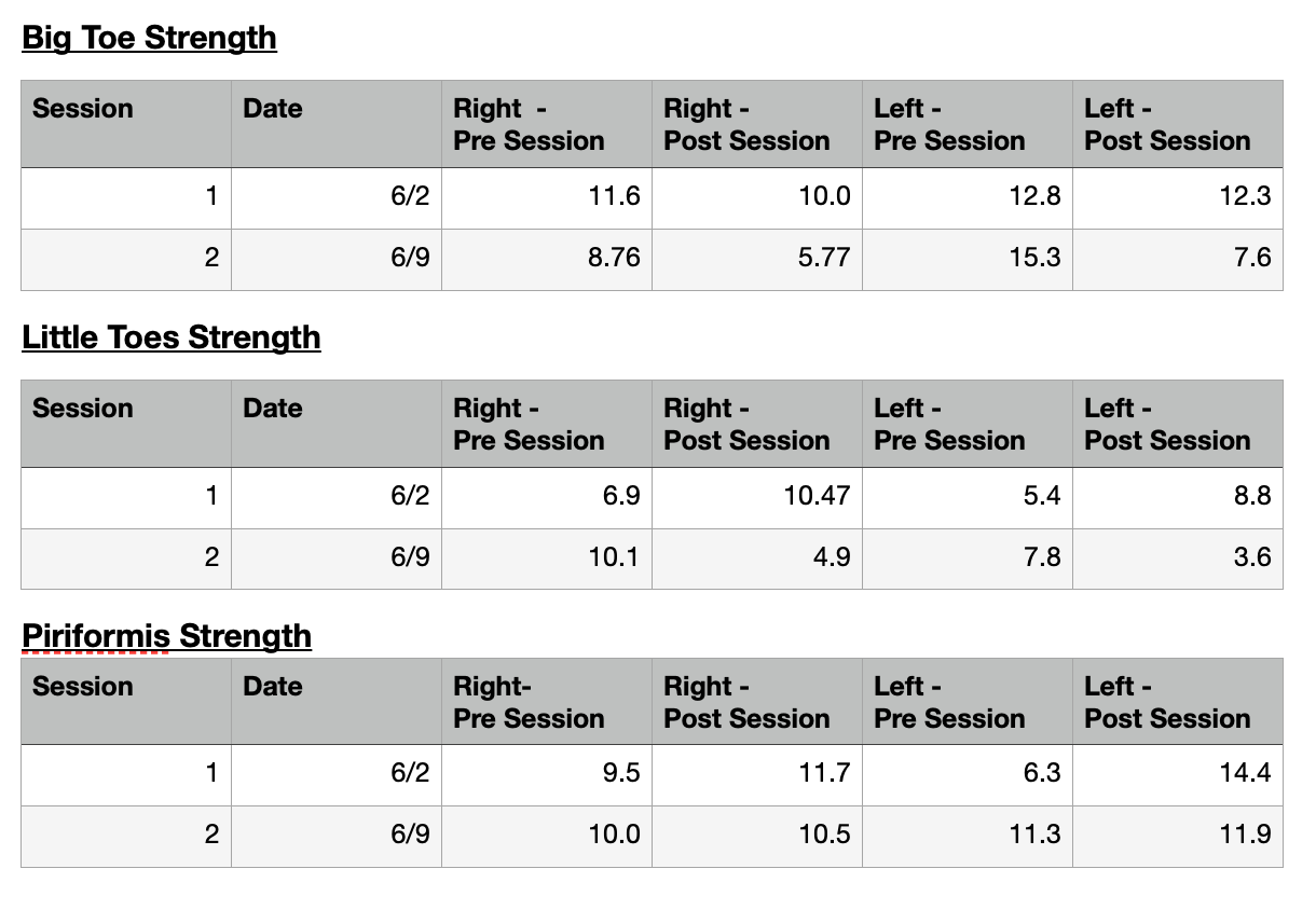
A targeted intervention plan was implemented, including specific Movement-Based NVR techniques and followed by dynamic full body movements. Data collected using a ToePro Strength Dynamometer on two separate dates (June 2nd and June 9th) measured big toe strength, little toes strength, and piriformis strength pre- and post-session.
The discussion asserts the X-pattern reflects fascial line imbalances in the Deep Front Line (DFL) and Spiral Line (SPL) and that arterial dysfunction contributes to muscle impairment. The acceleration of X-pattern correction after introducing Movement-Based NVR suggests its crucial role beyond movement alone in creating lasting structural change. The framework for this approach is based on the principle the body’s capacity must exceed demand for improvement and that Movement-Based NVR increases tissue capacity by releasing protective tension and strain. Functional full body movements were chosen for their integrative nature and used to solidify these gains.
The hypothesized outcomes for a positive effect on the X-pattern were: decreased big toe strength post-session, increased little toes strength post-session, and increased piriformis strength post-session. Additionally, it was theorized that between sessions, big toe strength should decrease, and little toes and piriformis strength would increase, indicating a more balanced anterior transverse arch of the foot and lasting strength changes due to the combined approach. Although initial session data showed some alignment with hypotheses regarding improved foot mechanics and piriformis strength, the collective data highlighted the body's resilient, non-linear adaptive nature.
The study's premature conclusion due to an external client injury underscores the need for extended investigation into NVR's long-term influence on complex postural patterns like genu valgus. This study aims to contribute to understanding Movement-Based NVR's potential in managing and positively affecting a common musculoskeletal presentations like genu valgus.
Introduction
This case study report explores the application and observed effects of Movement-Based Neurovascular Release (NVR) techniques coupled with functional movement, on an individual presenting with a common postural pattern of genu valgus, commonly referred as an X-pattern in the legs. Genu valgus is a postural reference characterized by the knees shifting towards the midline of the body while the ankles remain separated, often leading to altered biomechanics, pain, and functional limitations.
Movement-Based NVR is a specialized therapy approach that focuses on releasing tension and restrictions within the fascial, neural, and vascular systems. By addressing these intricate connections, NVR can improve tissue mobility, reduce pain, and restore optimal muscular function. With the addition of specific functional movement to capture and train the Nervous System and Muscular System with newly found mobility and ease.
This report aims to detail the specific Movement-Based NVR interventions and functional movement applied in this case, document the progression of the individual's condition, and analyze the observed changes in their genu valgus presentation and associated symptoms. Through this detailed examination, this researcher seeks to contribute to the understanding of Movement-Based NVR’s potential role in the conservative management of a common musculoskeletal presentation like genu valgus.
Case Presentation
The client began coming to the studio in September of 2023 in an effort to relieve knee pain from a right ACL tear several years ago. The client wanted to feel stronger, with increased endurance, and to eliminate the knee pain, increase core strength, and the X-pattern aesthetic. In April 2024, the client fractured her right foot and was in an orthopedic boot for 6 weeks post. According to the client, the X-pattern came about slowly, many years ago. There was not a significant or traumatic injury that caused the X-pattern presentation. Starting in December 2024, the sessions with the client began including movement-based NVR work. The knee pain had been taken care of; however the X-pattern aesthetic lingers. Knowing the X-pattern can cause residual pain, strain, and does not allow for optimal postural patterning or performance it was the goal of this researcher to derive a specific pathway for this long term goal. Although the goal may have been aesthetic, in nature, the strain from such pattern can have an unmeasurable effect on other tissues throughout the body. The ability to come out of this held pattern and distribute the body’s strain equitably throughout would result in a desired balance of efficiency and ease therefore optimizing performance.
The following data points were collected using a ToePro Strength Dynamometer:
It was decided to terminate this specific sequence of Movement-Based NVR and functional movement at an early date after the client reported persistent back pain from cleaning the playroom in a weighted vest which occurred between Session 1 and 2.
Management and Outcome
The following plan for release was created based upon the client’s postural pictures and known history. Although the client has had success with prior sessions of NVR and functional movement; the following specific plan was created and completed in its entirety in each session, to assess if additional and specific benefit could occur for the X-pattern.
Movement Based NeuroVascular Release
Occipital Artery Taffy Pull
Supraspinatous and SternoCloid Mastoid Hold
Omahyoid and Axillary Pin with Balloon Lift
Wilted Flower
Twisted Diaphragm with Long Leg Lever
Hip Erector Spinae Pin
Quadratus Lomborum and CircumFlex Femoral Artery Release
Piriformis Release
General Hip Release
Obterator/Bladder Hip Release
Hamstring Release - specific to Adductor Magnus and Minimus
Lateral Calf/Soleous Release
Anterior Tibialis/Extensor Digitorum Longus Differentiation
Tarsal Tunnel Gathering
Integrated Roll Down Seated
Integrated Roll Down Standing
Succeeding the Movement-Based NeuroVascular Releases the following Functional Movements were completed:
Functional Movement
Seated - Fingertip Isometric Head Presses Front/Back/Sides
Standing - Trip Around the Hip
Standing - Foot on 2 Bricks - Hip Hinge
Standing - Foot on 2 Bricks - Hip Hinge & Hip Rotation
Seated - Long Leg Lift over small ball
CoreAlign - Statue on Wheels
CoreAlign with Naboso Wedges - Statue on Wheels
CoreAlign in Releve` - Statue on Wheels
CoreAlign With Naboso Wedges - Statue on Wheels BackStroke
CoreAlign in Releve` - Statue on Wheels Backstroke
Discussion
An X-pattern leg presentation reveals specific postural imbalances, often stemming from dysfunction within the Deep Front Line (DFL) and Spiral Line (SPL), as understood through Anatomy Trains and Anatomy Trains Body Reading.
The DFL is the body's core support system. When it fails to function properly, there isn't always an immediate, obvious loss of function. However, insufficient support, balance, or proper tone in the DFL can lead to an overall shortening of the body and encourage collapse in the lumbo-pelvic core. This lays the groundwork for negative compensatory adjustments in all the other myofascial lines. These compensations can manifest in various ways, including ankle pronation or supination, knee rotation and medial shift, pelvic rotation, rib rotation on the pelvis, a lifted or anteriorly shifted shoulder, and head tilt, shift, or rotation.[1]
The SPL wraps the body in a double helix or spiral. This spiral assists in maintaining balance across all planes of movement. The SPL has a significant role in the X-patten as it specifically helps to determine knee tracking in walking. As with the DFL, the SPL also has an interwoven connection to many of the other myofascial lines. In commonality with the DFL compensation patterns can manifest in various ways; included but not limited to: ankle pronation/supination, knee rotation and medial shift, pelvic rotation on feet, one shoulder lifted or anteriorly shifted, and head tilt, shift, or rotation.[1]
With a clear understanding of the DFL and SPL's roles, a framework was developed to directly influence these lines. Studies show a connection between arterial dysfunction and muscle impairment, highlighting the importance of adequate blood flow to release strained areas.[2] Exercise itself offers both macrovascular and microvascular health benefits, and it's widely recognized that consistent physical activity throughout life can reduce the risk of falls, fractures, frailty, and a decline in quality of life. However, an input of movement alone was not enough to create the desired change needed to unwind the strongly held X-pattern. The client had been in a plateau and the desired goal of lasting long term structural changes was not occurring.
Viewing the body through the linear lens that utilizing the muscular system could solely create the desired optimized result is insufficient. It is necessary to work with an understanding of the body in terms of relationships and interconnections, and the dynamic system it is. This system will form a pattern of posture and movement from which injury or postural issues may arise. The capacity to tolerate a load of any movement dictates the maintenance of posture. When demand outweighs capacity, injury or compensation begins. Conversely, when capacity exceeds demand, performance is optimized, and improvement can occur. This relationship can be summarized as:
Capacity > Demand = Improvement of Physical Health & Optimized Performance
Capacity = Demand = Maintenance of Physical Health
Capacity < Demand = Degradation of Physical Health Injury/Compensation[3]
When Movement-Based NVR is utilized as a modality, we increase a muscle's capacity by releasing the protective pattern of tension and strain. It is then essential to ensure the muscle recognizes its new capacity to maintain it and continually work towards maintenance and optimized performance. Therefore, capacity can be greatly influenced with the inclusion of Movement-Based NVR.
Specific functional movement pieces were chosen to create a full-body integrated model. This approach is rooted in Sherrington’s Law of Irradiation, which states that "a muscle working hard recruits neighboring muscles to also work, and if those neighboring muscles are already involved, it amplifies their strength." This law explains how working one muscle can effectively "recruit" nearby muscles to assist, leading to improved and correct muscle recruitment and greater postural strength. This law is evident in the Piriformis measurement. This is particularly important as the release work performed through Movement-Based NVR allows for reduced strain/tone in muscle and the unbinding of muscles from its neighbors. Hence the muscle will have a new structure. Consequently, the new muscle architecture can contract and activate in concert, therefore helping the muscles "remember" their function within the newly adapted architecture of differentiation and tone. Additionally, Sherrington’s Law allows for full body movement change via recruitment rather than contract and release individually.
Based upon these theories, the following were the original hypothesized goals:
Big Toe Strength would decrease post session
Little Toes Strength would increase post session
Piriformis Strength would increase post session
In comparison Session 1 versus Session 2
Big Toe Strength Pre-Session 1 > Big Toes Strength Pre-Session 2
Little Toes Strength Pre-Session 1 < Little Toes Strength Pre-Session 2
Piriformis Strength Pre-Session 1 < Piriformis Strength Pre-Session 2
These hypothesized results would explain a positive effect on the X-pattern. If the theorized goals were met, the Anterior Transverse Arch of the foot would have then become more balanced as evidence of more equal strength data points of Big Toe versus Little Toes. In addition, if Sherrington’s Law applies to the approach of Movement-Based NVR with the addition of functional movement, then appropriate lasting and residual strength should be apparent across the two sessions in toe strength and piriformis strength change.
These hypothesized results would be a linear result, that can be depicted as an example of long-term study results would be Figure 1:

Rather, it is clear, the body is a resilient nonlinear system, as the body accepts multiple inputs into its system to create output.[4] The sum of those inputs equals the available capacity of the body. A simple illustration of this would be:
Input(Nervous + Arterial + Muscular Systems + etc.) = Capacity
It is important to note, the dynamic nonlinear relationship of the body does not produce a linear proportional effect, as Figure 1 suggests. A more accurate representation would be a graph with more curves and wiggles of effect[4].
An example depiction of such long-term study results would be Figure 2:

This case report demonstrates how inputs of Movement-Based NVR plus Functional Movement could have a positive influence on Genu Valgus. For this study, in summary:
Big Toe Strength DECREASED post Session 1
Big Toe Strength DECREASED post Session 2
Little Toe Strength INCREASED post Session 1
Little Toe Strength DECREASED post Session 2
Piriformis Strength INCREASED post Session 1
Piriformis Strength INCREASED post Session 2
In comparison Session 1 versus Session 2
Big Toe Strength Pre-Session 1 > Big Toe Strength Pre-Session 2
Little Toes Strength Pre-Session 1 < Little Toe Strength Pre-Session 2
Piriformis Strength Pre-Session 1 < Piriformis Strength Pre-Session 2
The values for the Anterior Transverse Arch and Piriformis were decreasing or increasing as hypothesized in Session 1. Big Toe and Piriformis strength decreased and increased, respectively, as hypothesized in Session 2. Little Toes Strength Session 2 values did not follow the hypothesized goals. This may be a product of the overriding resilient nonlinear relationship of the body. Therefore, these values do not represent an increase on Capacity. Further action and a longer term study would be needed to adequately determine the degree of positive trajectory with the X-pattern for this plan.
References
Myers, T. W.(2020). Anatomy Trains: Myofascial Meridians for Manual Therapists & Movement Professionals (4th ed.). Elsevier.
Dvoretskiy S, Lieblein-Boff JC, Jonnalagadda S, Atherton PJ, Phillips BE, Pereira SL. Exploring the Association between Vascular Dysfunction and Skeletal Muscle Mass, Strength and Function in Healthy Adults: A Systematic Review. Nutrients. 2020 Mar 7;12(3):715. doi: 10.3390/nu12030715. PMID: 32156061; PMCID: PMC7146456.
Spina, Andreo A., Functional Range Assessment - Systems Thinking, January 2023. PowerPoint Presentation.
Meadows, D. H.(2008). Thinking in Systems: A Primer, Chelsea Green Publishing.